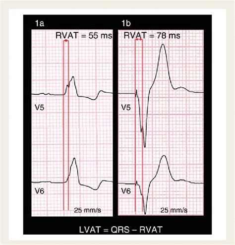
lv pacemaker lead placement | lead placement for heart failure lv pacemaker lead placement Although rare, inadvertent placement of a pacemaker or defibrillator lead in the left ventricle can have serious consequences, including arterial thromboembolism and aortic or mitral valve damage or infection. 1–4 By Jahla Seppanen January 4, 2022. Call it an understatement, but these are trying times. That being said, we need to find comfort where we can and one of the places that we can find comfort is in.
0 · pacing to left ventricular activation time
1 · pacing stimulus for left ventricular
2 · left ventricular lead resynchronization
3 · left ventricular lead placement cpt
4 · left ventricular lead placement
5 · lead placement for heart failure
6 · Lv lead placement in therapy
7 · Lv lead placement
$12K+
An optimal placement of the left ventricular (LV) lead appears crucial for the intended hemodynamic and hence clinical improvement. A well-localized target area and tools that help to achieve successful lead implantation seem to be of utmost importance to reach an optimal .
When the LBBP lead is used for cardiac resynchronization therapy (CRT) devices, the lead connection to the generator depends on the underlying rhythm (atrial fibrillation or sinus rhythm) and the choice of CRT-pacemaker or . The present article reviews the literature on image-guided cardiac resynchronization therapy (CRT) studies. Improved outcome to CRT has been associated with the placement of a left ventricular (LV) lead in the latest .Pacemaker lead placement through the tricuspid valve (TV) is infrequently associated with leaflet perforation and impingement of leaflet motion, resulting in valve dysfunction (Figures 10C and 10D) . When this leads to chronic fibrotic .
Although rare, inadvertent placement of a pacemaker or defibrillator lead in the left ventricle can have serious consequences, including arterial thromboembolism and aortic or mitral valve damage or infection. 1–4 Using an epicardial lead placed on the LV free wall via thoracotomy and endocardial leads placed in the right atrium (RA), left atrium (LA) via the coronary sinus (CS) .This is a video showing how to place an epicardial LV lead using the VATS technique. Learn more: https://www.ctsnet.org/article/vats-epicardial-lv-lead-place.It reaches the ventricle by penetrating the central fibrous body of the heart, where the fibres of left bundle branch (LBB) are given off after it emerges from the fibrous body at the level of the non .
Endocardial left ventricular (LV) pacing is an alternative therapy for patients who do not respond to conventional CRT or in whom placement of a lead via the coronary sinus is not .An optimal placement of the left ventricular (LV) lead appears crucial for the intended hemodynamic and hence clinical improvement. A well-localized target area and tools that help to achieve successful lead implantation seem to be of utmost importance to . CRT is a mainstay in the management of heart failure patients with electrical dyssynchrony. LV lead positioning remains an important variable that predicts response to CRT. Anatomical and technical challenges can hinder optimal LV lead placement using traditional lead implantation approaches.
When the LBBP lead is used for cardiac resynchronization therapy (CRT) devices, the lead connection to the generator depends on the underlying rhythm (atrial fibrillation or sinus rhythm) and the choice of CRT-pacemaker or CRT-defibrillator device. The present article reviews the literature on image-guided cardiac resynchronization therapy (CRT) studies. Improved outcome to CRT has been associated with the placement of a left ventricular (LV) lead in the latest activated segment free from scar.Pacemaker lead placement through the tricuspid valve (TV) is infrequently associated with leaflet perforation and impingement of leaflet motion, resulting in valve dysfunction (Figures 10C and 10D) . When this leads to chronic fibrotic changes in the TV, tethering of the leaflet often ensues. Although rare, inadvertent placement of a pacemaker or defibrillator lead in the left ventricle can have serious consequences, including arterial thromboembolism and aortic or mitral valve damage or infection. 1–4
Using an epicardial lead placed on the LV free wall via thoracotomy and endocardial leads placed in the right atrium (RA), left atrium (LA) via the coronary sinus (CS) and RV, they demonstrated a decrease in pulmonary capillary wedge pressure and an increase in cardiac output with temporary four-chamber pacing.
This is a video showing how to place an epicardial LV lead using the VATS technique. Learn more: https://www.ctsnet.org/article/vats-epicardial-lv-lead-place.It reaches the ventricle by penetrating the central fibrous body of the heart, where the fibres of left bundle branch (LBB) are given off after it emerges from the fibrous body at the level of the non-coronary aortic cusp. Endocardial left ventricular (LV) pacing is an alternative therapy for patients who do not respond to conventional CRT or in whom placement of a lead via the coronary sinus is not possible.
pacing to left ventricular activation time
An optimal placement of the left ventricular (LV) lead appears crucial for the intended hemodynamic and hence clinical improvement. A well-localized target area and tools that help to achieve successful lead implantation seem to be of utmost importance to . CRT is a mainstay in the management of heart failure patients with electrical dyssynchrony. LV lead positioning remains an important variable that predicts response to CRT. Anatomical and technical challenges can hinder optimal LV lead placement using traditional lead implantation approaches.
When the LBBP lead is used for cardiac resynchronization therapy (CRT) devices, the lead connection to the generator depends on the underlying rhythm (atrial fibrillation or sinus rhythm) and the choice of CRT-pacemaker or CRT-defibrillator device.
The present article reviews the literature on image-guided cardiac resynchronization therapy (CRT) studies. Improved outcome to CRT has been associated with the placement of a left ventricular (LV) lead in the latest activated segment free from scar.Pacemaker lead placement through the tricuspid valve (TV) is infrequently associated with leaflet perforation and impingement of leaflet motion, resulting in valve dysfunction (Figures 10C and 10D) . When this leads to chronic fibrotic changes in the TV, tethering of the leaflet often ensues. Although rare, inadvertent placement of a pacemaker or defibrillator lead in the left ventricle can have serious consequences, including arterial thromboembolism and aortic or mitral valve damage or infection. 1–4
Using an epicardial lead placed on the LV free wall via thoracotomy and endocardial leads placed in the right atrium (RA), left atrium (LA) via the coronary sinus (CS) and RV, they demonstrated a decrease in pulmonary capillary wedge pressure and an increase in cardiac output with temporary four-chamber pacing.This is a video showing how to place an epicardial LV lead using the VATS technique. Learn more: https://www.ctsnet.org/article/vats-epicardial-lv-lead-place.It reaches the ventricle by penetrating the central fibrous body of the heart, where the fibres of left bundle branch (LBB) are given off after it emerges from the fibrous body at the level of the non-coronary aortic cusp.
pacing stimulus for left ventricular
dillards michael kors wallets

cream michael kors wallet

left ventricular lead resynchronization
Experience sophistication at AX The Palace, a 5-star luxury hotel in Sliema offering both a city lifestyle as well as panoramic views of the Mediterranean Sea. Unlock up to 20% off your escape by booking directly through our website.
lv pacemaker lead placement|lead placement for heart failure


