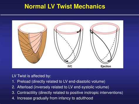
lv contraction | left ventricular contraction lv contraction The normal pattern of LV contraction has been defined as a uniform, concentric, inward motion of all points along the ventricular inner surface during systole. Uniform wall motion depends on . CONTACT US. Malts of Exceptional Quality. An incomparable process. Malting in the Eastern Townships, Quebec. Malting barley and floor malt made for microbrewery.
0 · lv twist mechanics
1 · left ventricular contraction normal range
2 · left ventricular contraction function
3 · left ventricular contraction
With ACE English Malta, finding accommodation options in Malta .
Late in diastole, atrial contraction increases the atrial pressure, producing a second atrial-to-LV pressure gradient that again propels blood into the LV. After atrial systole, as the left atrium .The normal pattern of LV contraction has been defined as a uniform, concentric, inward motion of all points along the ventricular inner surface during systole. Uniform wall motion depends on . The contraction of subepicardial fibers will rotate the apex of the LV in counterclockwise and its base in clockwise direction. Conversely, the contraction of .The compliance and resistance of the peripheral vascular system. The cardiac cycle has three defined phases: LV contraction. LV relaxation. LV filling. *Whilst similar phases are in .
The primary central abnormality in HF is an impaired left ventricular (LV) contractility or contractile reserve. Patients with an LV ejection fraction (LVEF) less than 50% (or <40% depending on .The triangular shape of RV PVL is explained by a shortened isovolumic contraction, an early peak chamber pressure, and an ESP close to the diastolic pressure, as ejection continues longer .
Many aspects of left ventricular function are explained by considering ventricular pressure–volume characteristics. Contractility is best measured by the slope, Emax, of the end . In routine clinical practice, assessment of left ventricular (LV) contractility is probably one of the most important tasks during evaluation of cardiac function.Left ventricular (LV) contraction dyssynchrony is common among patients with systolic heart failure and is associated with significantly greater cardiac risks. 1 Dyssynchronous contraction . Left ventricular contraction forces oxygenated blood through the aortic valve to be distributed to the entire body. With such an important role, decreased function caused by injury .
Late in diastole, atrial contraction increases the atrial pressure, producing a second atrial-to-LV pressure gradient that again propels blood into the LV. After atrial systole, as the left atrium .
lv twist mechanics
The normal pattern of LV contraction has been defined as a uniform, concentric, inward motion of all points along the ventricular inner surface during systole. Uniform wall motion depends on . The contraction of subepicardial fibers will rotate the apex of the LV in counterclockwise and its base in clockwise direction. Conversely, the contraction of .The compliance and resistance of the peripheral vascular system. The cardiac cycle has three defined phases: LV contraction. LV relaxation. LV filling. *Whilst similar phases are in .The primary central abnormality in HF is an impaired left ventricular (LV) contractility or contractile reserve. Patients with an LV ejection fraction (LVEF) less than 50% (or <40% depending on .
The triangular shape of RV PVL is explained by a shortened isovolumic contraction, an early peak chamber pressure, and an ESP close to the diastolic pressure, as ejection continues longer .
Many aspects of left ventricular function are explained by considering ventricular pressure–volume characteristics. Contractility is best measured by the slope, Emax, of the end .
In routine clinical practice, assessment of left ventricular (LV) contractility is probably one of the most important tasks during evaluation of cardiac function.
left ventricular contraction normal range
left ventricular contraction function
left ventricular contraction
Left ventricular (LV) contraction dyssynchrony is common among patients with systolic heart failure and is associated with significantly greater cardiac risks. 1 Dyssynchronous contraction . Left ventricular contraction forces oxygenated blood through the aortic valve to be distributed to the entire body. With such an important role, decreased function caused by injury .
Late in diastole, atrial contraction increases the atrial pressure, producing a second atrial-to-LV pressure gradient that again propels blood into the LV. After atrial systole, as the left atrium .
The normal pattern of LV contraction has been defined as a uniform, concentric, inward motion of all points along the ventricular inner surface during systole. Uniform wall motion depends on .
The contraction of subepicardial fibers will rotate the apex of the LV in counterclockwise and its base in clockwise direction. Conversely, the contraction of .The compliance and resistance of the peripheral vascular system. The cardiac cycle has three defined phases: LV contraction. LV relaxation. LV filling. *Whilst similar phases are in .The primary central abnormality in HF is an impaired left ventricular (LV) contractility or contractile reserve. Patients with an LV ejection fraction (LVEF) less than 50% (or <40% depending on .
The triangular shape of RV PVL is explained by a shortened isovolumic contraction, an early peak chamber pressure, and an ESP close to the diastolic pressure, as ejection continues longer . Many aspects of left ventricular function are explained by considering ventricular pressure–volume characteristics. Contractility is best measured by the slope, Emax, of the end . In routine clinical practice, assessment of left ventricular (LV) contractility is probably one of the most important tasks during evaluation of cardiac function.
hermes capucine vs orange

hermes birkin 25 black swift
Maisons à vendre - Malte. 109 annonces. Lieu exact. Les plus récentes. Elégante maison de ville rénovée avec piscine à Zabbar. Ħaż-Żabbar. Maison mitoyenne • 3 pce (s) • 3 .
lv contraction|left ventricular contraction


